A is for Airway: The Uncompromising Priority in Emergency Medicine
In the critical realm of emergency care, the sequence of actions is not arbitrary; it is a meticulously designed protocol to address the most immediate threats to life first. This protocol begins with the letter A, representing the Airway. An obstructed or compromised airway leads to oxygen deprivation, brain damage, and death within minutes. No other intervention, no matter how advanced, holds any value if oxygen cannot reach the lungs. The management of the airway is therefore the absolute foundation upon which all other emergency treatments are built, a complex skill set that ranges from simple manual maneuvers to advanced surgical procedures.
Anatomy of the Airway: A Pathway for Life
Understanding the airway’s structure is paramount to managing it effectively. The airway is divided into two main sections: the upper and lower airway.
The upper airway consists of the nasal cavity, the oral cavity, the pharynx (throat), and the larynx (voice box). Its primary functions are to warm, humidify, and filter inhaled air. The tongue is the most common cause of upper airway obstruction in an unconscious patient; as muscle tone decreases, the tongue falls back against the posterior pharynx, blocking the flow of air. The epiglottis, a leaf-shaped flap of cartilage, acts as a trapdoor that closes over the larynx during swallowing to prevent food and liquid from entering the lower airway.
The lower airway begins at the larynx, which contains the vocal cords and the narrowest part of the adult airway—the glottic opening. Below the larynx lies the trachea (windpipe), which further divides into the right and left main bronchi, branching into smaller bronchioles within the lungs. Protection of the lower airway from aspiration (inhalation of foreign material like vomit or blood) is a primary objective of advanced airway management.
The Spectrum of Airway Obstruction: Causes and Recognition
Airway compromise can be partial or complete, and it can occur at any point along its anatomical path. Causes are broadly categorized into mechanical, inflammatory, traumatic, and neurological.
- Mechanical Obstruction: This includes the tongue obstructing the pharynx in an unconscious patient, foreign body aspiration (e.g., food, a small toy), and severe trauma to the face or neck causing structural collapse or hematoma formation.
- Inflammatory/Infectious Obstruction: Conditions like anaphylaxis cause rapid swelling of the tongue, lips, and pharynx (angioedema). Infections such as epiglottitis (now rare due to vaccination) or peritonsillar abscess can also swell shut the airway.
- Traumatic Obstruction: Direct trauma from assaults, motor vehicle collisions, or burns can cause fractures, swelling, bleeding, and tissue damage that occludes the airway. Inhalation burns can cause rapid edema and swelling internally.
- Neurological Obstruction: A decreased level of consciousness from any cause (drug overdose, alcohol intoxication, head injury, stroke) leads to a loss of protective gag and cough reflexes, making the patient unable to maintain their own airway and highly susceptible to obstruction by their tongue or aspiration.
Recognizing airway obstruction is the first step. Signs can be obvious or subtle:
- Complete Obstruction: The patient cannot speak, cough, or breathe. They may universally clutch their neck (the universal choking sign) and rapidly become cyanotic (blue-tinged skin, especially around the lips and nail beds).
- Partial Obstruction: The patient may exhibit noisy breathing, a telltale sign of turbulent air moving through a narrowed passage.
- Snoring suggests tongue-based obstruction.
- Gurgling indicates the presence of liquid (blood, vomit, secretions) in the upper airway.
- Stridor is a high-pitched, harsh sound heard most often on inspiration, signaling a partial blockage at the level of the larynx or trachea.
- Wheezing is a whistling sound typically on expiration, indicating lower airway obstruction, as in asthma or COPD.
Other critical signs include agitation or confusion (early signs of hypoxia, or low oxygen) and lethargy or unconsciousness (late and dire signs of hypoxia and hypercarbia, or high carbon dioxide).
The Progression of Intervention: Basic to Advanced Maneuvers
Airway management follows a logical, stepwise progression, moving from simple, non-invasive techniques to complex, invasive procedures.
1. Basic Airway Maneuvers and Adjuncts:
The initial response to a suspected airway problem does not require any equipment.
- Head-Tilt/Chin-Lift Maneuver: For any unresponsive patient without suspected spinal injury, this is the primary action. By tilting the head back and lifting the chin, the anterior neck structures are stretched, which physically pulls the tongue away from the back of the throat.
- Jaw-Thrust Maneuver: This is the preferred initial maneuver for any patient with a suspected cervical spine injury. The rescuer places their fingers behind the angles of the patient’s mandible (jawbone) and displaces the jaw forward without moving the neck. This also pulls the tongue forward to open the airway.
- Suctioning: If gurgling sounds are present, immediate suctioning is required to clear blood, vomitus, or secretions. A large-bore, rigid suction catheter (Yankauer) is most effective for the oropharynx.
- Basic Adjuncts: These devices help maintain a patent airway without advanced skill.
- Oropharyngeal Airway (OPA): A curved plastic device that sits over the tongue, holding it anteriorly. It is only used in deeply unconscious patients without a gag reflex, as it can trigger vomiting or laryngospasm if inserted in a conscious or semi-conscious person.
- Nasopharyngeal Airway (NPA): A soft rubber tube that is passed through the nostril into the posterior pharynx. It is better tolerated in patients who have a diminished gag reflex but are not fully unconscious.
2. Advanced Airway Management:
When basic measures are insufficient to maintain a patent or protected airway, advanced techniques are necessary. This typically refers to endotracheal intubation.
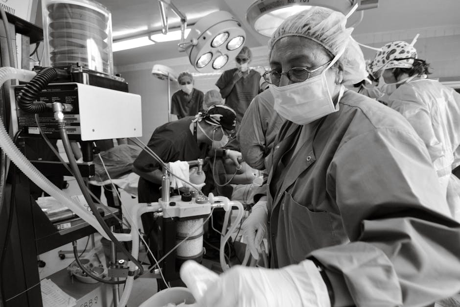
- Endotracheal Intubation (ETI): This is the gold standard for definitive airway control. It involves passing a cuffed tube through the vocal cords into the trachea. The cuff is then inflated to create a seal, which secures the airway, prevents aspiration, and allows for positive pressure ventilation. This procedure is performed using a laryngoscope to visualize the vocal cords or, increasingly, with a video laryngoscope, which provides a superior view and higher success rates. Confirmation of correct tube placement is absolutely critical and is verified by multiple methods: auscultation of breath sounds over both lungs and the stomach, observation of chest rise, condensation in the tube, and, most reliably, by using a quantitative carbon dioxide (CO2) detector (capnography).
3. Rescue and Surgical Options:
When attempts at securing an airway through intubation fail, a “can’t intubate, can’t oxygenate” scenario arises, which is a life-threatening emergency. Rescue options include:
- Supraglottic Airways: Devices like the laryngeal mask airway (LMA) are inserted blindly into the pharynx, forming a seal over the laryngeal inlet. They are easier to place than an ET tube and serve as a critical rescue device or a temporary airway.
- Surgical Cricothyrotomy: This is a last-resort, invasive procedure performed when all other methods have failed. It involves making an incision through the skin and the cricothyroid membrane (located between the thyroid cartilage and the cricoid cartilage) to place a tube directly into the trachea. It provides a definitive, though temporary, airway to oxygenate the patient.
Special Considerations and Pitfalls
Airway management is not a one-size-fits-all process. Specific scenarios demand tailored approaches.
- Pediatric Airways: Children are not small adults. Their airways are anatomically different: a larger tongue relative to the mouth, a higher and more anterior larynx, a narrower trachea, and the narrowest part of the airway is at the cricoid cartilage (not the vocal cords). These differences make them more susceptible to obstruction and more challenging to intubate.
- C-Spine Precautions: In any patient with potential trauma, the cervical spine must be immobilized manually throughout all airway maneuvers to prevent exacerbating a spinal cord injury. The jaw-thrust maneuver is essential here.
- Rapid Sequence Intubation (RSI): This is a structured pharmacologic technique used in patients who have a gag reflex but still require a definitive airway. It involves the simultaneous administration of a sedative (induction agent) and a neuromuscular blocking agent (paralytic) to quickly render the patient unconscious and paralyzed, facilitating intubation while minimizing the risk of aspiration and combating the patient’s own physiology.
- The Pitfall of Oxygenation vs. Ventilation: A critical concept is that oxygen can be moved into the lungs of a patient with a completely obstructed airway via passive diffusion if a tight seal is made with a high-concentration oxygen mask. This provides a few precious extra minutes to perform a definitive procedure like a cricothyrotomy. It underscores the principle that while securing the airway is paramount, oxygenation is the ultimate goal.
The Role of Continuous Monitoring and Capnography
Securing the airway is only the beginning. Continuous monitoring is essential to ensure it remains secure. The tube can become dislodged, kinked, or obstructed. The most important technological advancement in this monitoring is continuous waveform capnography. This device measures the concentration of exhaled CO2 and displays a waveform. A consistent waveform is the gold standard confirmation that the endotracheal tube is placed in the trachea and remains there. Any change in the waveform or CO2 level provides an immediate early warning of a problem, such as esophageal intubation, tube dislodgement, or a physiological change in the patient’s status like a drop in cardiac output. It is an indispensable tool for patient safety.